

 Previous page
Image analysis is a powerful tool used to solve microscopic problems but
it also can be dangerous when used in an automated capacity. We have used
the image analysis computer program Diana to describe crystal size and
vesicle size distributions in Mt. St. Helens gray dacite (MSHD) and Merapi
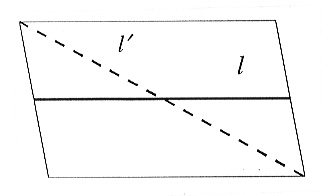
basaltic andesite (MRP9603), first using the length of the crystal to characterize
its size. The program considers the longest dimension of the crystal as
its length, irrespective of its crystallographic axes, causing both errors
in true length (defined by a crystallographic axis) and crystal orientation
(Fig. 3.8-7). An improved analysis is made using the area of the crystals
as size compared to the short axis. We found the results of the later method
consistent with the crystal accumulation in the particle-distribution of
experimentally obtained pyroclasts of MRP9603. While using the method on
pyroclast samples of Mt. St. Helens gray dacite, the distribution curve
did not verify our image analytical results. In determining the crystal-distribution
Diana did not recognize a fracture system in the phenocryst. Closer examination
of the dacite using reflected light revealed that most of the phenocrysts
were broken. In MSHD the mechanism that hinders fracturing from destroying
the phenocrysts failed to do so in MRP9603. SEM images of MSHD experimental-pyroclastic
particles show multiple pyroclasts (glass and
crystal) in all size fractions, whereas MRP9603 reveals many unbroken crystals
with thin glass rims.
 |
|
|
Characterizing vesicle sizes in pyroclastic rocks is commonly carried out by the examination of binary images of analyses. Either scanned SEM or reflection-light-microscopy images are used for the investigations. Most primary (non-filtered) pictures are composed of a wide range of grayscales (256). Transferring the images into binary files, the program searches for a selected grayscale to calculate the black and white border. Diana allows some recalculation in the event of illumination effects from light microscopy. Materials like MSHD ground mass do not allow precise measurement by reflected light microscopy, and SEM pictures are required. By microscopic investigation we found two vesicle-systems in MSHD: a few large (mm) irregularly formed vesicles and a network of several small micron-wide tubes. A 2-D image analysis of these branched tubes shows that 34% of the MSHD-ground mass is built up of vesicles with a 3 µm diameter. BET (multi-point gas sorption) surface analysis reveals a surface area of 0.1347m²/g for MSHD, or 2.36 m2 for the 17.5 g sample analyzed. We then calculate the open tube system of 3 µm diameter to have a length of approximately 24000 m. To describe this system on a 2-D basis is not possible. Mercury immersion tests have revealed a third locally connected vesicle system of micron size. In order to use image analysis to describe this type of vesicle system we will correlate permeability measurements and vesicle-form-characteristics as is practiced by sedimentologists.

Tel: +49-(0) 921 55 3700 / 3766, Fax: +49-(0) 921 55 3769, E-mail: bayerisches.geoinstitut(at)uni-bayreuth.de